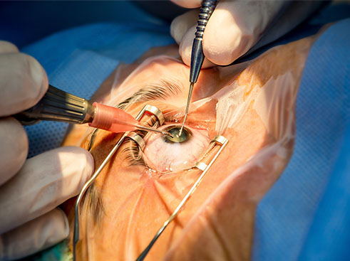
Management of Vitreous Loss in Cataract Surgery
This course will increase your confidence during cataract surgery by providing tools to mitigate the complication of vitreous loss.

CONTEXT
Vitreous loss is the most common and feared complication of cataract surgery. In surveys of ophthalmologists who have attended vitrectomy courses, it is evident that this is a leading cause of concern for most ophthalmologists.
This anxiety associated with vitreous loss is based on the fact that many anterior segment surgeons have not had adequate training in vitrectomy.
This course hopes to provide the fundamentals and recommendations to prevent or solve this complication.
Therefore, a vitreous loss course for anterior segment surgeons is necessary. All the knowledge, techniques, and strategies that vitreous surgeons have developed are used and adapted to the situation of limbal surgery as well.
THE DETAILS
Intended for: Ophthalmologists, ophthalmologists in training, subspecialists in various areas of ophthalmology.
Duration: Approximately six weeks, 2-3 hours a week.
Certificate: 12 hours of PAAO Continuing Medical Education on completion
OBJECTIVES
At the end of the course, the participants will be able to:
- Determine the measure of risk of vitreous loss.
- Compare different surgical techniques and what the indications are according to the findings.
- Describe the most common vitrectomy equipment’s functions, parameters, and connection systems and make the connections.
- Prematurely recognize the signs of vitreous loss.
- Define the principles of a well-done vitrectomy and apply them to all situations in which a complication occurs.
- List and describe the different intraocular lens insertion techniques in the event of capsular rupture or loss of capsular support.
- Describe the intraocular complications that occur as a result of vitreous loss that is not appropriately managed.
PROGRAM
- Vitreous loss: introduction to the course and justification. Statistics of vitreous loss and its consequences. When traditional management is performed, what is the pathophysiology of vitreous loss, and why is it possible to avoid damage with an adequate vitrectomy.
- Determination of risk in cataract surgery. A detailed description of the 21 variables that intervene in the definition of risk. A simplified method to determine which patients should be made a risk table.
- Description of a typical vitrectomy. What does a vitrectomy do? How to define the suction and cut variables. Commonly used vitrectomy connections: Infiniti, Constellation, EVA, Zeiss. Fluidics in vitrectomy, work cycle. Tips 20, 23, 25, and 27. Use of endo illumination. Use of the 76 diopter lens and contact lenses with limbal illumination. Pedal configuration and variables on screen.
- Alternatives to Phacoemulsification. How should extracapsular surgery be done in high-risk cataracts? Remaining indications for intracapsular surgery. When to guide the patient for pars plana phaco fragmentation. Training and level of expertise required for each of these options. How to perform vitrectomy in the presence of a wide incision.
- Early signs of vitreous loss. The importance of early recognition of capsular rupture. Management alternatives according to the stage of surgery. How and when to convert to extracapsular. Measures to avoid vitrectomy in case of capsular rupture. Capsular expansion rings and the importance of their early use. Variants of sutured rings.
- Vitreous loss in 4 moments of the surgery: when the phaco is starting, in the aspiration of debris, when the capsule is being polished, and after inserting the intraocular lens. Technical variants in each situation.
- Insertion of a 3-piece IOL, prolene loops, wide total diameter, when there is a central capsular rupture and preservation of the capsular rim. Lens options must be available in every surgery. Artisan IOL insertion for inverted aphakia when there is no capsular support. Scleral fixation IOL insertion with aniridia lens option with loops sutured to the sclera. Kenzo-Yamame technique for scleral fixation of prolene loops.
- The consequences of vitreous loss are when it is not managed properly, respecting the principles set forth or when they are not followed.
COURSE MATERIALS
- Study guides, that facilitate the student’s learning process.
- Audiovisual lectures prepared by the teaching team on each topic. These will be available for online reading and in a save/print version.
- Reading material, available in Spanish and/or English in pdf format.
- Concept maps
LEARNING ACTIVITIES
Learning activities used throughout this course:
- Multiple choice questions.
- Open questions, self-corrected with checklists.
- Question and answer forums.
Each module will have an evaluation. The number of attempts is unlimited.
The activities will promote the exchange and use of students’ knowledge and experiences and facilitate the application of new learning to professional practice.
Meet the faculty

Dr Alberto Castro
Course Director

Dr. Eduardo Mayorga
Instructional Design
PAAO Member
Resident/Fellow
$100 USD
$120 USD non-member
life-time access
forum support
printable study materials
PAAO Member
Ophthalmologist
$150 USD
$200 USD non-member
life-time access
forum support
printable study materials
Ophthalmic
Technician*
$200 USD
USD non-member
life-time access
forum support
printable study materials
**Technicians must submit a letter from an ophthalmologist.

